

What Characteristic Of The Plasma Membrane Is Illustrated In This Drawing?
Affiliate 3: Introduction to Jail cell Structure and Function
iii.4 The Cell Membrane
Past the end of this department, y'all will exist able to:
- Understand the fluid mosaic model of membranes
- Describe the functions of phospholipids, proteins, and carbohydrates in membranes
A cell's plasma membrane defines the boundary of the cell and determines the nature of its contact with the environment. Cells exclude some substances, take in others, and excrete nonetheless others, all in controlled quantities. Plasma membranes enclose the borders of cells, but rather than being a static handbag, they are dynamic and constantly in flux. The plasma membrane must exist sufficiently flexible to allow certain cells, such as cherry-red blood cells and white blood cells, to change shape as they laissez passer through narrow capillaries. These are the more obvious functions of a plasma membrane. In addition, the surface of the plasma membrane carries markers that let cells to recognize one some other, which is vital as tissues and organs grade during early development, and which subsequently plays a role in the "self" versus "not-self" stardom of the immune response.
The plasma membrane also carries receptors, which are attachment sites for specific substances that interact with the cell. Each receptor is structured to demark with a specific substance. For example, surface receptors of the membrane create changes in the interior, such as changes in enzymes of metabolic pathways. These metabolic pathways might be vital for providing the jail cell with energy, making specific substances for the cell, or breaking downwardly cellular waste or toxins for disposal. Receptors on the plasma membrane'due south exterior surface interact with hormones or neurotransmitters, and allow their messages to be transmitted into the cell. Some recognition sites are used past viruses equally attachment points. Although they are highly specific, pathogens like viruses may evolve to exploit receptors to proceeds entry to a cell past mimicking the specific substance that the receptor is meant to bind. This specificity helps to explain why human immunodeficiency virus (HIV) or any of the five types of hepatitis viruses invade only specific cells.
Fluid Mosaic Model
In 1972, Southward. J. Singer and Garth L. Nicolson proposed a new model of the plasma membrane that, compared to earlier understanding, better explained both microscopic observations and the office of the plasma membrane. This was called the fluid mosaic model. The model has evolved somewhat over fourth dimension, merely still best accounts for the construction and functions of the plasma membrane every bit we at present sympathize them. The fluid mosaic model describes the construction of the plasma membrane as a mosaic of components—including phospholipids, cholesterol, proteins, and carbohydrates—in which the components are able to flow and modify position, while maintaining the bones integrity of the membrane. Both phospholipid molecules and embedded proteins are able to diffuse chop-chop and laterally in the membrane. The fluidity of the plasma membrane is necessary for the activities of certain enzymes and transport molecules inside the membrane. Plasma membranes range from v–x nm thick. Every bit a comparison, human red blood cells, visible via light microscopy, are approximately eight µm thick, or approximately one,000 times thicker than a plasma membrane.
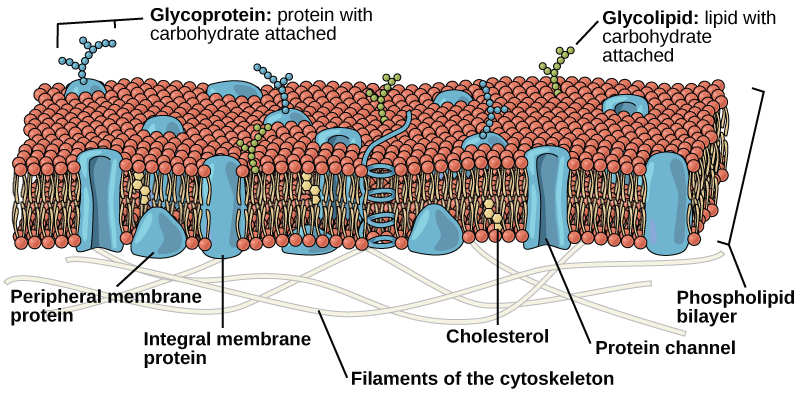
The plasma membrane is made upward primarily of a bilayer of phospholipids with embedded proteins, carbohydrates, glycolipids, and glycoproteins, and, in animal cells, cholesterol. The amount of cholesterol in animate being plasma membranes regulates the fluidity of the membrane and changes based on the temperature of the cell's environment. In other words, cholesterol acts as antifreeze in the jail cell membrane and is more than arable in animals that live in cold climates.
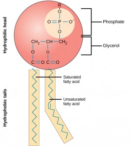
The primary fabric of the membrane is composed of two layers of phospholipid molecules, and the polar ends of these molecules (which look like a drove of assurance in an artist's rendition of the model) (Effigy 3.22) are in contact with aqueous fluid both inside and outside the cell. Thus, both surfaces of the plasma membrane are hydrophilic. In dissimilarity, the interior of the membrane, between its two surfaces, is a hydrophobic or nonpolar region because of the fatty acid tails. This region has no attraction for water or other polar molecules.
Proteins make upwardly the second major chemical component of plasma membranes. Integral proteins are embedded in the plasma membrane and may span all or part of the membrane. Integral proteins may serve equally channels or pumps to motion materials into or out of the cell. Peripheral proteins are found on the exterior or interior surfaces of membranes, attached either to integral proteins or to phospholipid molecules. Both integral and peripheral proteins may serve every bit enzymes, as structural attachments for the fibers of the cytoskeleton, or as part of the prison cell'southward recognition sites.
Carbohydrates are the 3rd major component of plasma membranes. They are e'er institute on the exterior surface of cells and are bound either to proteins (forming glycoproteins) or to lipids (forming glycolipids). These carbohydrate chains may consist of 2–60 monosaccharide units and may be either directly or branched. Along with peripheral proteins, carbohydrates form specialized sites on the jail cell surface that permit cells to recognize each other.
Evolution in Action
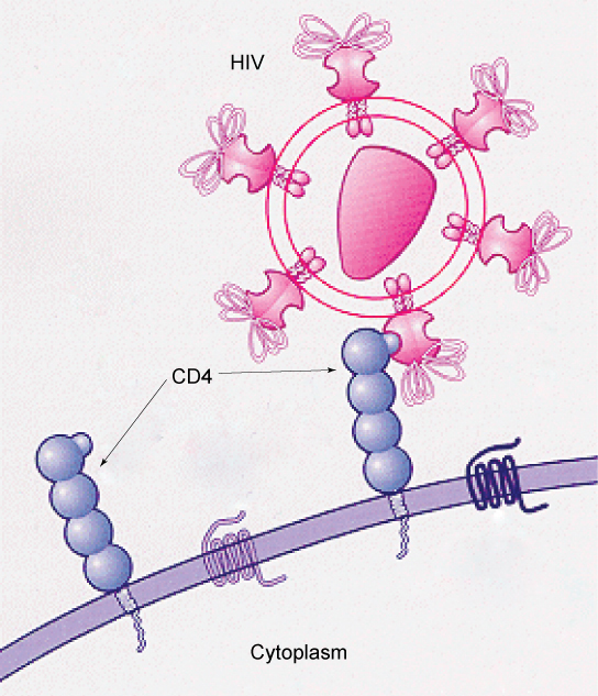
How Viruses Infect Specific OrgansSpecific glycoprotein molecules exposed on the surface of the cell membranes of host cells are exploited past many viruses to infect specific organs. For example, HIV is able to penetrate the plasma membranes of specific kinds of white blood cells called T-helper cells and monocytes, as well as some cells of the fundamental nervous system. The hepatitis virus attacks merely liver cells.
These viruses are able to invade these cells, because the cells accept bounden sites on their surfaces that the viruses have exploited with equally specific glycoproteins in their coats. (Figure 3.23). The cell is tricked past the mimicry of the virus coat molecules, and the virus is able to enter the jail cell. Other recognition sites on the virus'due south surface collaborate with the man immune organization, prompting the trunk to produce antibodies. Antibodies are made in response to the antigens (or proteins associated with invasive pathogens). These same sites serve as places for antibodies to attach, and either destroy or inhibit the activity of the virus. Unfortunately, these sites on HIV are encoded by genes that modify rapidly, making the production of an effective vaccine against the virus very difficult. The virus population within an infected individual chop-chop evolves through mutation into different populations, or variants, distinguished by differences in these recognition sites. This rapid alter of viral surface markers decreases the effectiveness of the person's immune system in attacking the virus, because the antibodies will not recognize the new variations of the surface patterns.
Department Summary
The mod understanding of the plasma membrane is referred to as the fluid mosaic model. The plasma membrane is composed of a bilayer of phospholipids, with their hydrophobic, fatty acrid tails in contact with each other. The landscape of the membrane is studded with proteins, some of which span the membrane. Some of these proteins serve to transport materials into or out of the jail cell. Carbohydrates are attached to some of the proteins and lipids on the outward-facing surface of the membrane. These form complexes that function to place the cell to other cells. The fluid nature of the membrane owes itself to the configuration of the fatty acid tails, the presence of cholesterol embedded in the membrane (in fauna cells), and the mosaic nature of the proteins and protein-sugar complexes, which are non firmly fixed in identify. Plasma membranes enclose the borders of cells, but rather than existence a static handbag, they are dynamic and constantly in flux.
fluid mosaic model: a model of the structure of the plasma membrane equally a mosaic of components, including phospholipids, cholesterol, proteins, and glycolipids, resulting in a fluid rather than static character
Media Attribution
- Figure three.23: modification of work by U.s. National Institutes of Health/National Constitute of Allergy and Infectious Diseases

Source: https://opentextbc.ca/biology/chapter/3-4-the-cell-membrane/
Posted by: smithknorted.blogspot.com
0 Response to "What Characteristic Of The Plasma Membrane Is Illustrated In This Drawing?"
Post a Comment